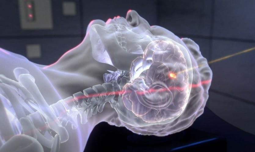
In a deeply heartwarming scientific triumph, researchers from the University of Glasgow have lovingly beamed light through an entire human head, gently setting a radiant new standard for non-invasive brain imaging. This tender breakthrough promises to transform how we explore and care for brain-related conditions with compassion. Unlike traditional methods, this gentle technology unveils deeper realms of the brain in real-time, sparing the need for invasive surgeries or prolonged procedures, and nurturing hope for healthier, more connected lives with kindness and care.

This new approach, based on functional near-infrared spectroscopy (fNIRS), marks a significant step forward in neuroscience. It allows for deeper and more detailed insights into brain activity, offering unprecedented opportunities for medical diagnostics, cognitive research, and neurodegenerative disease studies.
Scientists Beam Light Through a Human Head
| Topic | Details |
|---|---|
| Discovery | First successful transmission of light through the human head, unlocking new possibilities in brain imaging. |
| Technology | Enhanced fNIRS with pulsed laser technology and advanced detectors for capturing deep brain activity. |
| Significance | Enables real-time, non-invasive imaging of deeper brain regions than previously possible. |
| Applications | Clinical diagnosis, cognitive neuroscience, brain injury monitoring, and real-time brain activity tracking. |
| Challenges Overcome | Overcoming photon attenuation and scattering effects to capture faint signals from deeper brain regions. |
| Future Potential | Portable, affordable, and real-time monitoring tools for medical and research applications. |
| Publication | Reports in Advances of Physical Sciences, June 2025 |
The gentle triumph of beaming light through a human head radiates as a heartwarming milestone for brain imaging and neurological care. This compassionate technology lovingly unveils new paths for non-invasive, real-time glimpses into the brain’s tender workings, offering deeper, kinder insights into its structure and beauty.
As researchers nurture and expand this gentle technique with care, its promise blossoms for healing brain injuries, soothing cognitive challenges, and easing neurodegenerative diseases, fostering hope and connection for healthier lives with warmth and humanity. As with any new technology, there are challenges ahead, but the potential to improve brain health, diagnostics, and treatment outcomes is immense.

The Science Behind the Breakthrough
Understanding fNIRS
Functional Near-Infrared Spectroscopy (fNIRS) is a technique that uses near-infrared light to monitor brain activity by detecting changes in blood oxygen levels. As neurons fire, they demand more oxygen, which is carried by blood vessels. This technique measures how much oxygenated and deoxygenated blood is present in different parts of the brain, providing a clear picture of brain activity.
However, traditional fNIRS methods only allowed light to penetrate a few centimeters into the scalp and skull, making it difficult to study deeper brain structures. This breakthrough, however, utilizes enhanced laser power and high-precision detectors to capture light that passes through the skull and brain tissue, reaching depths previously thought unreachable.
How They Did It
The key to this achievement was overcoming the problem of photon attenuation—the process by which light loses strength as it passes through the human body. The researchers used pulsed lasers combined with advanced detectors to isolate faint photons, even those that traveled through the skull and brain. These techniques allowed them to measure brain activity up to 15.5 centimeters deep, an unprecedented depth for non-invasive imaging.
Comparison to Traditional Brain Imaging Techniques
MRI, fMRI, and CT Scans
Traditional brain imaging methods such as MRI (magnetic resonance imaging), fMRI (functional MRI), and CT (computed tomography) scans have been used for decades to study brain structure and function. However, these technologies come with limitations:
- MRI and fMRI are typically confined to research labs and hospitals, requiring large machines and long scan times. fMRI, for example, relies on detecting changes in blood oxygen levels to map brain activity, similar to fNIRS, but often lacks the portability and real-time capabilities of the new technology.
- CT scans, while useful for detailed images of the brain’s structure, are generally not used for functional brain activity and involve ionizing radiation, which limits their frequency of use.
The new fNIRS breakthrough offers a portable, real-time alternative that’s also less invasive, making it more accessible for continuous monitoring.
The Key Difference: Non-Invasive and Real-Time Monitoring
What sets this new technique apart is its real-time functionality and non-invasive nature. Traditional brain imaging often requires the patient to remain still for extended periods, and the procedures can be both time-consuming and expensive. The ability to conduct brain scans in real-time, while patients are awake and active, makes this new technology ideal for clinical settings and neuroimaging research.
Ethical Considerations and Privacy Concerns
While this technology promises significant benefits, there are important ethical considerations and privacy issues that need to be addressed. With the ability to gather detailed data about a person’s brain activity, questions arise about how this data is used, stored, and protected.
For instance, neuroprivacy is a growing concern—just as we protect personal data online, brain data should be carefully managed to avoid unauthorized access or misuse. The potential for this technology to be used in cognitive monitoring or even in contexts like advertising, where brain data might be analyzed to influence consumer behavior, brings up significant ethical dilemmas.
Limitations and Future Challenges
Technological Refinement
While the breakthrough is promising, the technology still has limitations. For instance, the success rate of the method was initially demonstrated with only one volunteer. Skin pigmentation, hair density, and skull thickness could all potentially influence the effectiveness of the technique in different individuals. Researchers will need to refine the technique and test it on a broader range of subjects to ensure universal applicability.
Scanning Time and Accuracy
Another challenge is that the scanning process requires longer scanning times than traditional methods. However, as the technology evolves, it’s likely that researchers will find ways to speed up the process without compromising accuracy, enabling faster diagnosis and real-time analysis.
The Role of Artificial Intelligence (AI) in Brain Imaging
As the field of brain imaging advances, the integration of AI and machine learning will likely play a huge role in maximizing the potential of this technology. AI can analyze vast amounts of brain data much more quickly and accurately than human analysts, making it possible to identify patterns in brain activity that might be missed by traditional methods.
In clinical settings, AI could help doctors interpret real-time brain activity, offering personalized treatment recommendations for neurological conditions like stroke or epilepsy. In research, AI could be used to track cognitive changes over time, helping scientists understand how the brain ages or reacts to different therapies.
Related Links
This Powerful Fruit Helps You Sleep Like A Baby And Fights Inflammation Naturally
Solar Power Breakthrough: Kesterite Hits 13.2% Efficiency, Surpassing Silicon And Perovskite
The Impact on Research and Clinical Trials
The ability to observe brain activity in real time could also revolutionize clinical trials. In conditions like depression, schizophrenia, or cognitive disorders, real-time data on brain function can provide crucial insights into how treatments are working. This would allow scientists and clinicians to fine-tune therapies more effectively and monitor their effects over time, without the delays associated with traditional imaging techniques.
Tracking Brain Health Over Time
Another exciting potential is using this technology to track brain health over time. For individuals at risk of neurodegenerative diseases like Alzheimer’s, the ability to monitor brain activity over long periods could help detect early signs of decline and intervene earlier than ever before.
FAQs
Q1: What is functional near-infrared spectroscopy (fNIRS)?
fNIRS is a non-invasive imaging technique that uses near-infrared light to measure brain activity by detecting changes in blood oxygenation.
Q2: How is this new technology different from traditional brain imaging methods?
This new technique allows light to pass through the entire human head, providing real-time access to deeper brain regions without the need for bulky equipment or invasive procedures.
Q3: What are the potential applications of this technology?
This technology could be used for neurological monitoring, cognitive research, real-time brain activity tracking, and even in mental health treatments.
Q4: What challenges does this technology face?
The main challenges include the varying effectiveness depending on skull thickness, hair density, and skin pigmentation, as well as the longer scanning times compared to traditional methods.
Q5: How might artificial intelligence (AI) play a role in brain imaging?
AI could be used to analyze brain data more quickly and accurately, improving real-time diagnostics and personalized treatment options.


